Sports Injury Physiotherapy
Experts in sporting recovery
At The Spinal Physiotherapy & Sports Medicine Clinic we have a team of highly trained and skilled Chartered Physiotherapists who have years of experience working across a wide variety of sports, all the way from pitch side at your local underage team to international and Olympic competition in sports such as Horse Racing, Rowing, Golf, Football, Rugby, Athletics, Ice Hockey, Gaelic Football, Badminton, Cycling, Fencing….pretty much everything you can think of.
Muscle Strain
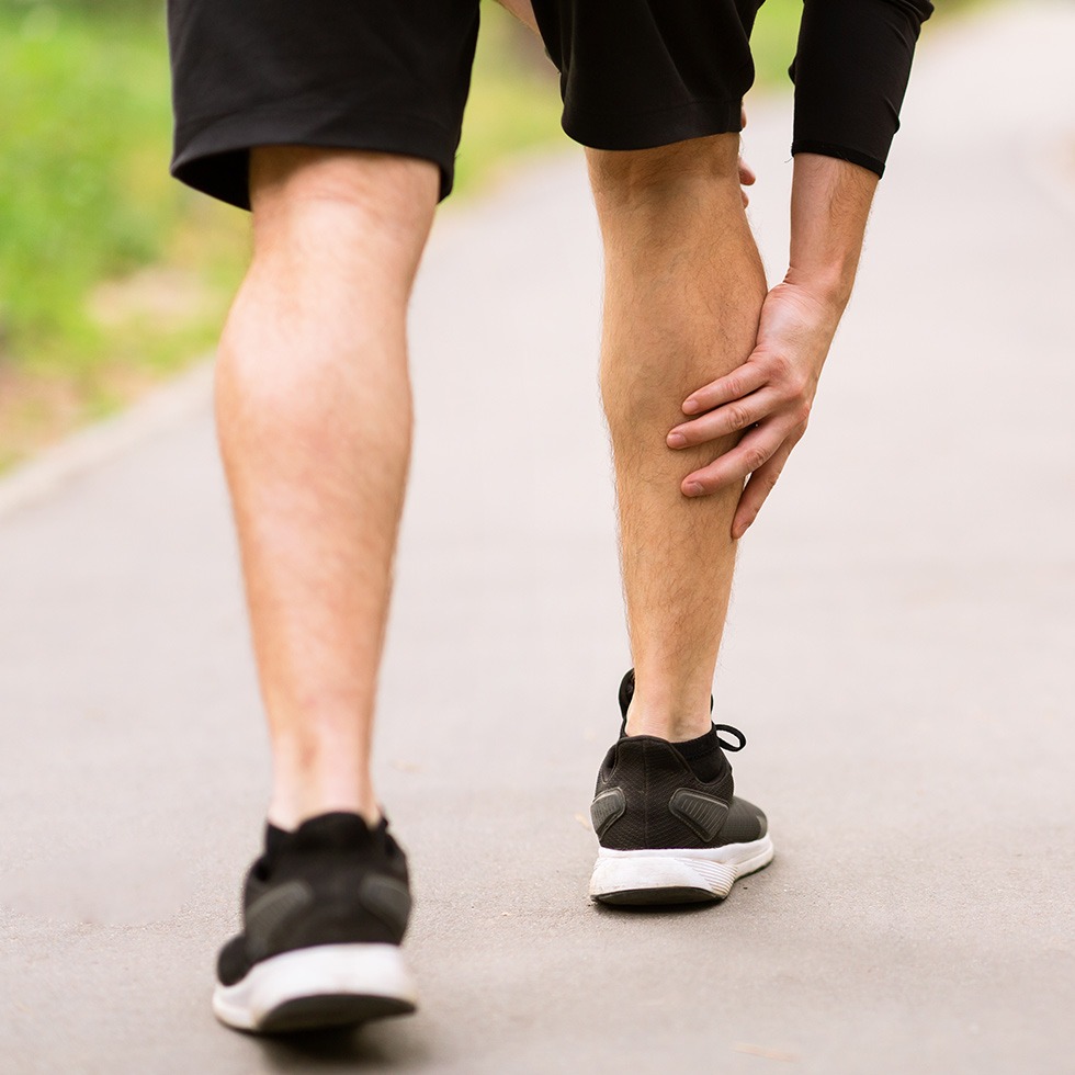
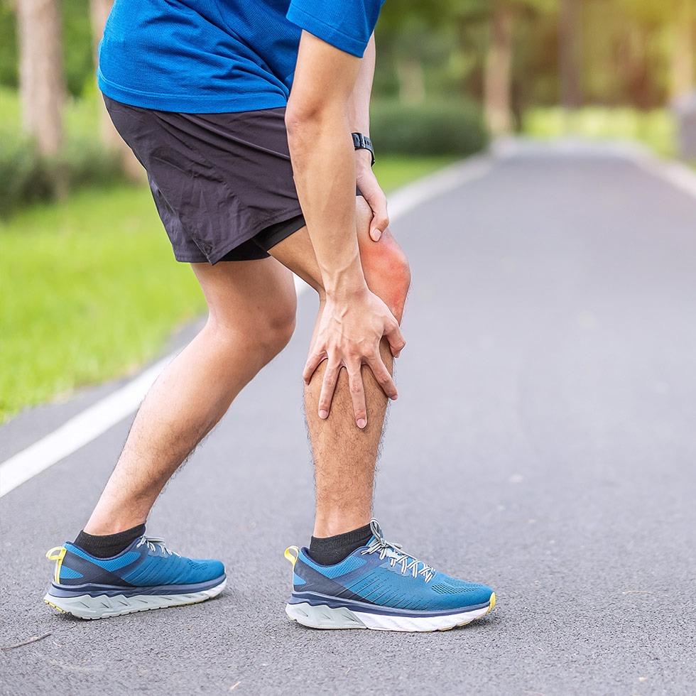
A muscle strain or ‘pulled muscle’ occurs when the muscle belly or the tendon which attaches the muscle to bone, is overstretched or torn. Eccentric contractions (the muscle contracting while it is being stretched) can cause this overstretching or tearing.
A muscle strain is a common sports injury. Running, kicking and jumping are actions which frequently result in a muscle strain. An improper warm-up before activity; weak muscles; overuse and insufficient recovery time are reasons why a muscle can become strained. Lifting a heavy object using an improper lifting technique is also a common cause.
Strains are graded on a scale indicating the severity of the injury. A grade I muscle strain is mild, the muscle has been overstretched; it has not torn. The muscle may feel tender and slightly stiff, but a grade I strain should not affect function greatly.
A grade II strain is more severe as the muscle is partially torn. The muscle will feel stiff and painful and the area may be swollen and bruised. Trying to use the muscle will be painful and therefore a grade II strain will affect function.
A grade III strain is the most severe and means the muscle has been completely torn. You will have no muscle function. There may be a dent present over the muscle area and it will very painful and swollen.
Following an assessment, a physiotherapist will be able to diagnose the grade of muscle strain you have sustained.
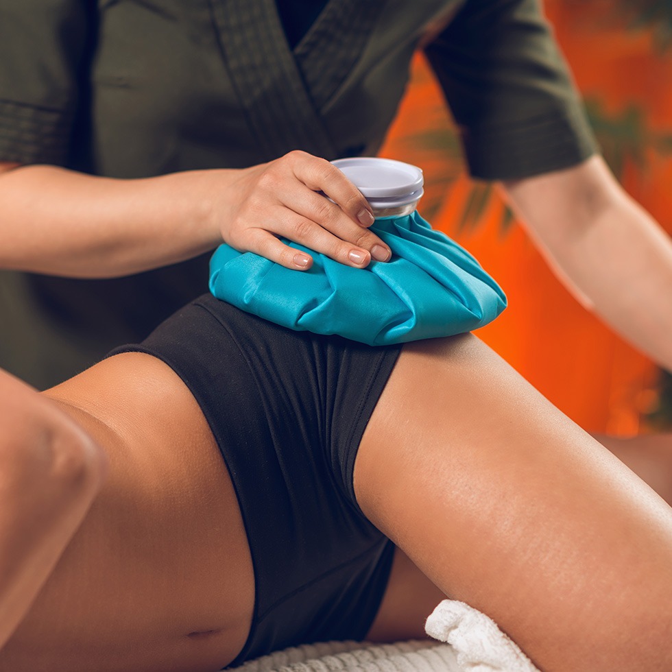
After any muscle strain it is important to protect the area from further damage. You should rest the affected area for the first 2-3 days following injury, refraining from substantial activity. Applying ice to the muscle can reduce swelling and bruising if used during the first 2-3 days following injury. Ice should not be applied directly to the skin. Place a cloth between the icepack/ bag of frozen peas to prevent an ice burn. Apply the ice for approximately 20 minutes every few hours and, if appropriate, elevate the area. Also, swelling can be reduced by applying compression using an elasticated tubular bandage. Ensure this is not too tight and never wear overnight. Keeping the affected area elevated as much as possible will help reduce any swelling and therefore help reduce pain. It is beneficial to the healing process to begin gentle movements 3 days after your injury as long as they are relatively pain free.
What kind of physiotherapy you receive after a muscle strain will depend on the severity of the strain. A physiotherapist will provide appropriate treatment to regain range of movement, strength and flexibility at the affected area. Undergoing proper rehabilitation after a muscle strain is important in order to reduce the risk of reoccurrence of injury.
Ligament Sprain
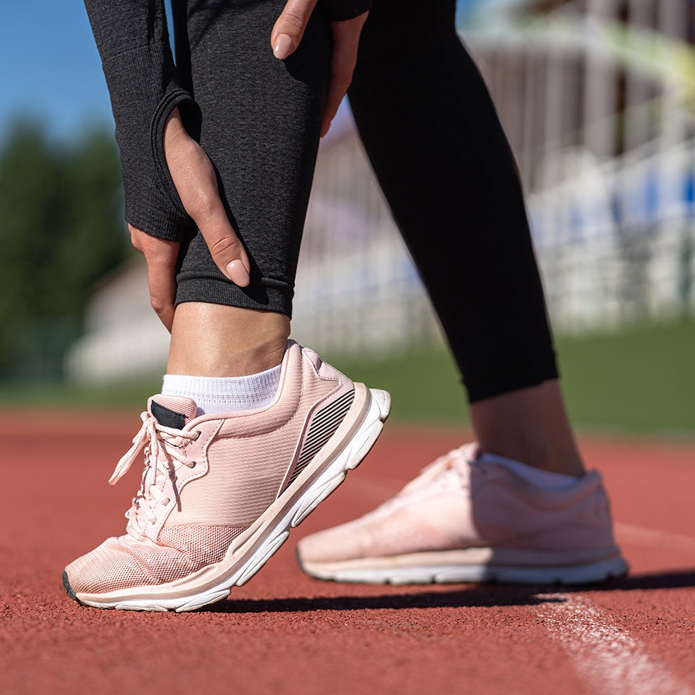
Ligaments connect bone to bone and help control the range of movement in a joint to keep it within a stable range. Ligaments also have numerous sensors called proprioceptors that feed back to the brain to let us know where we are in time and space.
If the force through a ligament is so great that it causes the joint to go beyond its normal range then the ligament will break down, resulting in a sprain. Sprains have three grades.
- Grade I – A small number of collagen fibres in the ligament are damaged resulting in an inflammatory response with local tenderness over the ligament. Can often be treated with good advice and self management.
- Grade II – A significant number of fibres within the ligament are damaged causing a larger inflammatory response, increased swelling and intense pain. Physiotherapy is required for these injuries.
- Grade III – Complete rupture of the ligament. Large amount of swelling, instability of the joint and intense pain. May require surgery to restore the stability to the joint
Tendinopathy
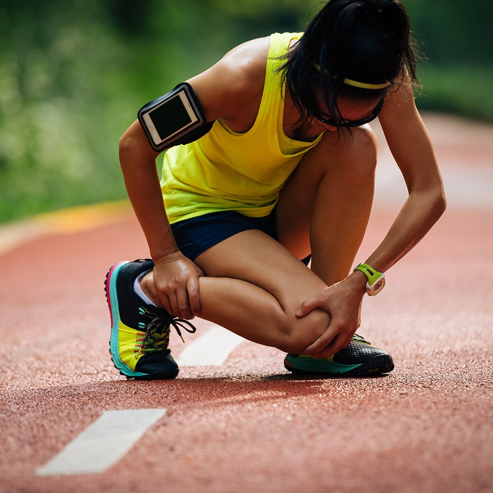
The term “itis” means inflammation and we now tend to use the word tendonopathy for a problem with a tendon as it isn’t always clear if the tendon is actually inflammed or not. Either way what ultimately happens is that the tendon gets over loaded through repitive activity or poor control of that activity. The tendon will get small microdamage or tears as a consequence. If the rate of these microtears is greater than the rate of recovery, this will lead to a prolonged inflammatory phase known as tendonitis. That will give way to the tendon scarring or degenerating and this then becomes a tendonopathy, as the tendon isn’t necessarily inflammed anymore but there is pain, stiffness and limited ability to load the tendon to perform tasks.
The physiotherapists at spinal physio will be able to diagnose tendonopahties but more importantly identify the mechanisms as to why it developed and treat accordingly.

Knee Cartilage Injury
A torn cartilage is not an uncommon injury in sport. The knee has 2 discs of fibrocartilage called menisci. They are C-Shaped and slightly thicker at the edge of the joint in order to cushion to opening and closing mechanism of the knee.
The menisci are shock absorbers so will innevitably wear over time and lead to micro tears which can cause pain or discomfort and stiffness in the joint but can usually be managed conservatively with physiotherapy
More acute tears to the menisci occur in twisting injuries such as football, tennis, skiing and even golf. Imagine your meniscus is like a finger nail that you tear a bit off. You can tear a small piece and you don’t either notice or miss it. You can tear a larger piece that can stick up and isn’t much of a problem unless you catch it on something and then it will hurt. As the knee is a finite space that piece of cartilage that sticks up, sometimes referred to as a bucket handle tear can get caught in the joint like a biro in the hinge of a door and the knee just won’t move. This is called locking of the knee. This requires surgery to remove the piece of cartilage and is normally done with key hole surgery or an arthroscopy. You can also tear off a reasonably large part of the cartilage that floats around the knee, known as a loose body, which can sometimes cause locking or maybe not at all.
If you have locking of the knee, chances are you need surgery. If you have a stiff, painful, swollen or tender joint but no locking or giving way after a twisting injury you will probably require and benefit from physiotherapy.
Patellofemoral Pain
The patella is the floating bone over the knee and the femur is the thigh bone. The patello-femoral joint is the joint between these two structures.
The patella is an egg shaped bone sitting on top of the knee joint. It’s slightly rounded at the top and pointed at the bottom. The quadriceps muscles are 4 muscles from the hip that all converge on the patella and result in a single tendon, the patellar tendon connecting to the Tibia or shin.
Picture the patella like a wheel in a pulley and the quadriceps are 4 muscles or ropes going into the pulley with one rope, the patellar tendon coming out the other side. If everything is pulling evenly then the wheel of the pulley will operate smoothly and the hinge of the knee works very well. If the ropes pull at different rates or strengths as a result of muscle imbalance, then the patella will tilt and this will cause friction or rubbing of the patella on surrounding structures.
The physiotherapists at spinal physio will be able to assess your knee to establish the dirtection of deviation as this can be medially, laterally, anteriorly, superiorly, inferiorly or any combination of the above. Depending on the mal-tracking will determine which structures are being irritated and what treatment and/or exercise program is required.
It is important with patellofemoal dysfunction to not only treat the structures that have been damaged by the mal-tracking but also the tracking issue itself and this may involve the stability around the hip and pelvis, possibly the back or feet and biomechanics. It’s simple really.
Jumpers Knee
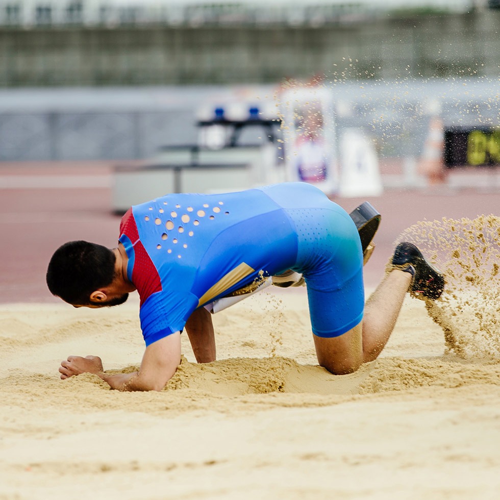
Patellar tendonitis is otherwise known as jumpers knee. The term “itis” means inflammation and we now tend to use the word tendonopathy for a problem with a tendon as it isn’t always clear if the tendon is actually inflammed or not. Either way what ultimately happens is that the tendon gets over loaded through repitive activity or poor control of that activity. The tendon will get small microdamage or tears as a consequence. If the rate of these microtears is greater than the rate of recovery, this will lead to a prolonged inflammatory phase known as tendonitis. That will give way to the tendon scarring or degenerating and this then becomes a tendonopathy, as the tendon isn’t necessarily inflammed anymore but there is pain, stiffness and limited ability to load the tendon to perform tasks.
The physiotherapists at spinal physio will be able to diagnose patellar tendonopahties but more importantly identify the mechanisms as to why it developed and treat accordingly.
Femeroacetabular Impingement
The hip is a ball and socket joint. It is made up of the ball on top of the thigh bone called the femur and the socket in the pelvis known as the acetabulum. The socket of the hip is surrounded by a layer of fibrocartilagenous material known as the labrum. You can tear the labrum just like a torn cartilage in the knee. See the Knee cartilage injury section below.
Femeroacetabular impingement is as it suggestes. An impingement between the femur or ball of the hip and Acetabulum or socket of the hip. This can have a number of forms but there are two main types. A picer deformity and a Cam type deformity.
A Pincer deformity is where the ends of the socket are more pointed and pincer like making the sockey more C-Shaped than the subtle U shape it should be. This can then mean that the ball of the hip rubs on one of the pincer ends causing impingement and pain.
A Cam type deformity is where the ball of the hip becomes slightly more square like over time and you now have a square peg trying to fit into a round hole. This again will cause impingement and pain.
As with everything in anatomy, physiology and injury these conditions can be further classified into more specific types and the physiotherapists at Spinal Physio will be able to assess your hip to establish which issue you may have and how best to treat it.
Tennis Elbow
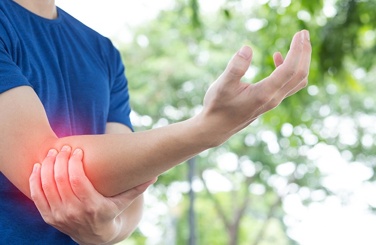
The tendons that extend or straighten our fingers all come from the same point of the elbow. This area is known as the lateral epicondyle or extensor tendon origin. Pain in this region is often described as lateral epicondylitis or tennis elbow. There are a number of tendons all originating from the same point and moving out into different directions to get to the fingers. As these tendons overlap and are working to move the hand and fingers particularly in gripping activities they can rub against each other creating friction, inflammation and eventually become scarred.
The tendon that moves the middle finger sits on top of all the other tendons and is the one that can rub the most. This tendon is called Extensor Carpi Radialis Brevis. In tennis elbow it is this tendon that is affected. There are other tendons that can be affected or other areas of pain on the lateral aspect of the elbow, however it is not tennis elbow unless the middle finger is involved.
Classically the tendon will be rubbing and slowly fraying for months. It usually takes about 6 months for the process to lead to pain or inflammation. Tennis elbow is characterized by pain in the lateral or outer aspect of the elbow. The pain often originates behind the elbow joint and radiates out from there over the outer upper one third of the forearm. It is intermittent at first and gradually deteriorates becoming increasingly stiff and painful. Most patients present with symptoms occurring for approximately 3 months at which time the tendon has become scarred and very weak. In all they have had the problem for 9 months by this time.
It is necessary to establish the source of the problem. With careful questioning a source is usually identified which more than likely will involve an increase in gripping activity in the previous 6-9 months. Classic examples are a short intense spell of DIY, gardening, gripping sports such as tennis or golf or perhaps a lot of right clicking with the mouse using the middle finger.
This intense period of activity may have started the scarring process and although not painful at the time, slowly degenerates resulting in the pain of tennis elbow 6 months later. If the cause can be identified this will greatly improve the chances of recovery.
Physiotherapy can conservatively manage tennis elbow and will result in full pain free and functional recovery in 70-80% of cases. The physiotherapist will break down the scar tissue and start the patient on a graduated exercise program. It will take 12 weeks to fully recover and the patient will need on average 8 sessions in that time.
The physiotherapist may consider a brace to help reduce the pain during treatment. Most of these are ineffective. The best tennis elbow supports are those that stop the hand from moving rather that restrict elbow function as it is gripping of the hand that is responsible for the damage in the first instance. The physiotherapist may also try acupuncture to control the pain during treatment.
If physiotherapy is unsuccessful the next option is to have one or a series of cortisone injections. There is much debate over the effectiveness of cortisone in tennis elbow as there are many who believe that there is no inflammation. There is also debate as to how many and where exactly these injections should be administered. The general consensus is that 3 injections would be the maximum over a 6 month period.
Surgery is the final resort and this will either involve debridement of the scar tissue or a tendon transfer. Tendon transfers are the more successful surgical procedure as it changes the angle at which the tendon pulls. This relieves the stress on the tendon and should reduce it’s recurrence in the future. Post surgical rehabilitation with a Physiotherapist is essential for good long term results.
Plantarfascitis
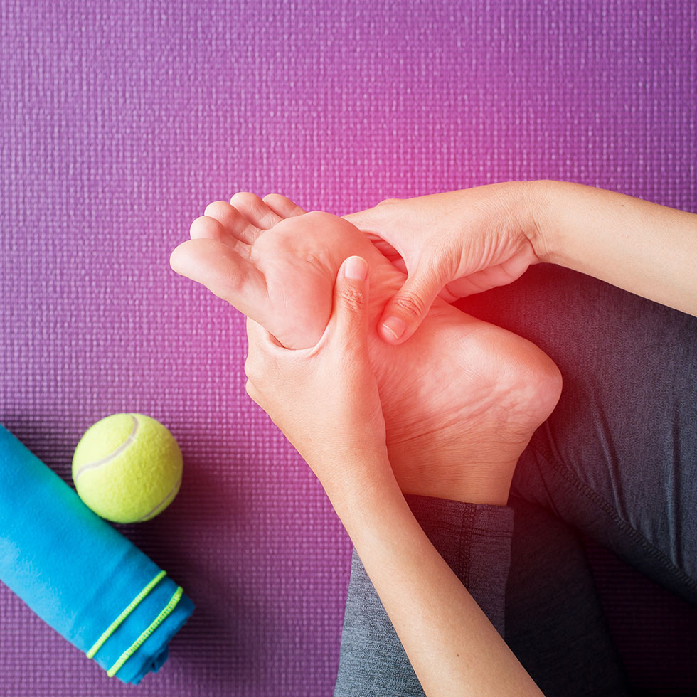
Plantar fasciitis is an inflammatory condition of the plantar facia, a thick band of fibrous tissue which runs along the sole of the foot from the heel to the toes. Continued overstretching of the plantar fascia can lead to micro tearing at the point where the plantar fascia inserts into the heel. This micro tearing can cause an inflammatory reaction in the local area, resulting in heel pain.
There are a number of reasons for the overstretching of the plantar fascia which can in turn lead to plantar fasciitis. These include foot arch problems, obesity, a tight Achilles tendon, wearing unsupportive footwear, and standing/walking on hard surfaces for long periods of time.
A possible symptom of plantar fasciitis is a heel spur (bony growth) which may eventually develop as a result of continued overstretching of the plantar fascia and associated ‘pulling’ at the insertion into the heel. A heel spur is not the cause of plantar fasciitis and is not what causes the heel pain. The vast majority of heel spurs grown parallel to the heel bone so don’t get in the way and don’t cause any problems. It is now considered pointless X-raying the heel looking for a heel spur as most that exist are insignificant anomalies. A very small number of heel spurs do grow vertically downwards and can be a source of pain but an examination by a physiotherapists will be able to inform you if an x-ray is appropriate.
If suffering from plantar fasciitis you will have pain on the underside of the heel which is often worse in the morning, during the first steps of the day. This sharp pain usually reduces to a dull ache throughout the day and reduces with rest, but may be quite painful to get going again after long periods of sitting down. In some cases the pain can also spread to the arch of the foot. The inflammation at the heel will also cause mild swelling, occasional redness, and it may be difficult to walk due to pain.
Applying ice to the local area can help reduce inflammation. Ice should not be applied directly to the skin. Place a cloth between the icepack/ bag of frozen peas to prevent an ice burn. Apply the ice to an elevated heel for approximately 20 minutes every few hours. Avoiding substantial activity and resting as much as possible will also help reduce inflammation and therefore the heel pain.
Following an assessment, a physiotherapist will be able to diagnose plantar fasciitis and provide the appropriate treatment, which may include periosteal pecking, orthotics and the provision of specific exercises and advice on how to prevent recurrence.
Online Consultation
We understand it’s not always possible to get to the clinic or you just have some questions that you’d like to discuss with an expert and get some advice.
With an online consultation you can have a 30 minute Zoom/Skype appointment with an experienced Chartered Physiotherapist who will discuss your concerns and offer appropriate advice.
